

Introduction
The human body, with all its intricacies, never ceases to amaze. Among the many organ-specific conditions that have puzzled researchers and clinicians alike, Gallbladder Adenomyomatosis stands out as an intriguing topic.
The gallbladder, though small, plays a pivotal role in our digestive system. As such, understanding any abnormalities related to it is crucial.
What is Gallbladder Adenomyomatosis?
Firstly, Gallbladder Adenomyomatosis, often simply referred to as Adenomyomatosis, is a benign, non-cancerous change in the gallbladder’s wall. When it occurs, the muscle layer of the gallbladder wall thickens and forms small pouches called diverticula.
These pouches may contain bile, and they protrude into the inner layer of the gallbladder. This condition is often discovered accidentally during imaging studies for other issues, and most patients are asymptomatic.
Causes and Risk Factors
The exact cause of Gallbladder Adenomyomatosis remains unknown. However, several theories suggest that it may result from increased gallbladder pressure or inflammation. These factors could cause the mucosa to herniate into the muscular layer, leading to the formation of the diverticula.
Risk factors might include:
- Chronic gallbladder inflammation
- Gallstones
- Age, as it’s more commonly found in older adults
Despite these associations, it’s essential to remember that having one or more risk factors does not mean you’ll develop the condition. Nevertheless, it’s always a good idea to be aware and seek regular check-ups, especially if you’re in the high-risk group.
Symptoms and Diagnosis
Interestingly, most people with Adenomyomatosis experience no symptoms at all. However, if symptoms do appear, they may resemble those of other gallbladder diseases. Common symptoms can include:
- Abdominal pain, particularly in the upper right quadrant
- Nausea or vomiting
- Bloating
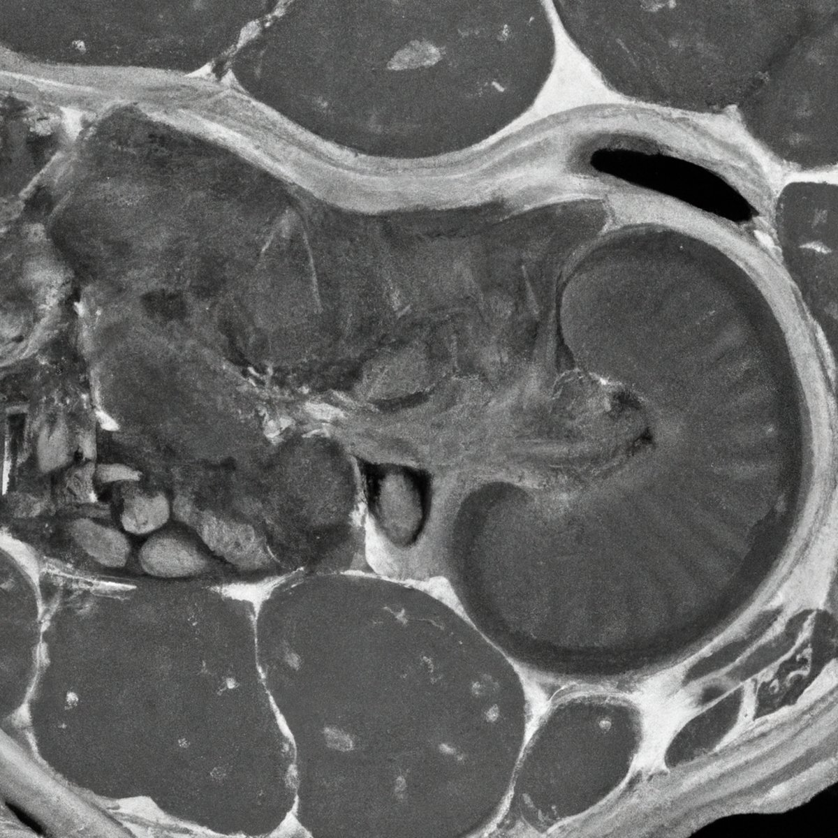
For diagnosis, doctors typically rely on imaging studies such as ultrasounds, MRIs, or CT scans. Because this condition often looks similar to other gallbladder diseases on scans, it requires an experienced radiologist to distinguish Adenomyomatosis accurately.
Treatment Options
Fortunately, Adenomyomatosis doesn’t always require treatment. Yet, if the patient is symptomatic or there’s a concern about the potential for gallbladder cancer, the physician might recommend surgery, usually a cholecystectomy (removal of the gallbladder).
It’s worth noting that the removal of the gallbladder is a standard procedure and the human body can function normally without it. Bile, which the gallbladder stores, will instead flow directly from the liver to the small intestine.
Living with Gallbladder Adenomyomatosis
If you’ve been diagnosed with this condition and are asymptomatic, there’s often little to worry about. However, one should maintain regular doctor visits to monitor the situation.
For those who have had their gallbladders removed due to Adenomyomatosis, life post-surgery is typically normal. Initially, you might have to adjust your diet to lower-fat meals, as the bile flow will now be continuous rather than stored for large meals.
Conclusion
In essence, Gallbladder Adenomyomatosis, although less commonly discussed, provides a compelling topic in the world of gastroenterology. The journey of understanding this condition underscores the complexity and wonder of the human body. By shedding light on such topics, we empower ourselves with knowledge, fostering better decision-making and peace of mind.
Always consult with a healthcare professional if you have concerns related to your gallbladder or any other health issues. Remember, every discovery and understanding in medical science begins with curiosity and the thirst for knowledge.