

Introduction
Xanthogranulomatous Cholecystitis (XGC) is a rare and potentially serious inflammatory condition that affects the gallbladder. While it is not as common as other gallbladder conditions, understanding its symptoms and diagnosis is crucial for timely intervention and treatment.
In this comprehensive article, we will delve into the world of XGC, exploring its symptoms and how medical professionals diagnose this condition.
What is Xanthogranulomatous Cholecystitis?
Before we dive into the symptoms and diagnosis, let’s first understand what Xanthogranulomatous Cholecystitis is. XGC is a rare form of cholecystitis characterized by the infiltration of the gallbladder wall by inflammatory cells, including foamy histiocytes, lymphocytes, and plasma cells. This infiltration results in the formation of yellowish nodules within the gallbladder tissue, which gives the condition its distinctive name.
XGC often occurs as a complication of chronic cholecystitis and is associated with gallstones. The exact cause of XGC is not well understood, but it is believed to be related to chronic inflammation and infection.
Symptoms of Xanthogranulomatous Cholecystitis
Abdominal Pain and Tenderness
One of the hallmark symptoms of XGC is persistent and severe abdominal pain and tenderness. This pain is usually located in the right upper quadrant of the abdomen, where the gallbladder is situated. Patients may describe the pain as sharp, stabbing, or aching. The discomfort can be continuous or intermittent but tends to worsen over time.
Jaundice and Yellowing of the Skin
Jaundice, the yellowing of the skin and the whites of the eyes, is another significant symptom of XGC. This occurs because the inflammation in the gallbladder can lead to a blockage in the common bile duct, preventing the normal flow of bile. As a result, bilirubin, a yellow pigment, accumulates in the body, causing the characteristic yellowing of the skin and eyes.
Fever and Chills
Fever and chills are common symptoms of any inflammatory process, including XGC. As the body’s immune system responds to the inflammation in the gallbladder, it often leads to an elevated body temperature. Patients may experience sweating and chills as their body tries to combat the infection.
Nausea and Vomiting
Nausea and vomiting can be a result of the pain and inflammation associated with XGC. Additionally, when the bile flow is obstructed due to the condition, it can lead to digestive disturbances, causing nausea and occasional vomiting.
Weight Loss and Fatigue
Unexplained weight loss and fatigue are less common but still important symptoms of XGC. These symptoms can occur as a consequence of the chronic inflammation, which affects overall health and nutrition.
Diagnosis of Xanthogranulomatous Cholecystitis
Diagnosing Xanthogranulomatous Cholecystitis is a multi-step process that involves a combination of clinical evaluation, imaging studies, and laboratory tests.
Physical Examination and Medical History
A crucial first step in the diagnosis of XGC is a thorough physical examination and a review of the patient’s medical history. The doctor will inquire about the patient’s symptoms, their duration, and any relevant medical conditions or risk factors.
Imaging Tests
Imaging tests are essential for confirming the presence of XGC. These tests may include:
Ultrasound: A non-invasive test that uses sound waves to create images of the gallbladder. It can show signs of inflammation and gallstones.
CT Scan: A computed tomography (CT) scan provides detailed cross-sectional images of the gallbladder and surrounding structures, helping to visualize any abnormalities.
MRI: Magnetic resonance imaging (MRI) can provide detailed images of the gallbladder and is especially useful for evaluating soft tissue changes.
Blood Tests
Blood tests are performed to assess the patient’s overall health and look for signs of infection or inflammation. Elevated white blood cell counts and abnormal liver function tests may suggest XGC.
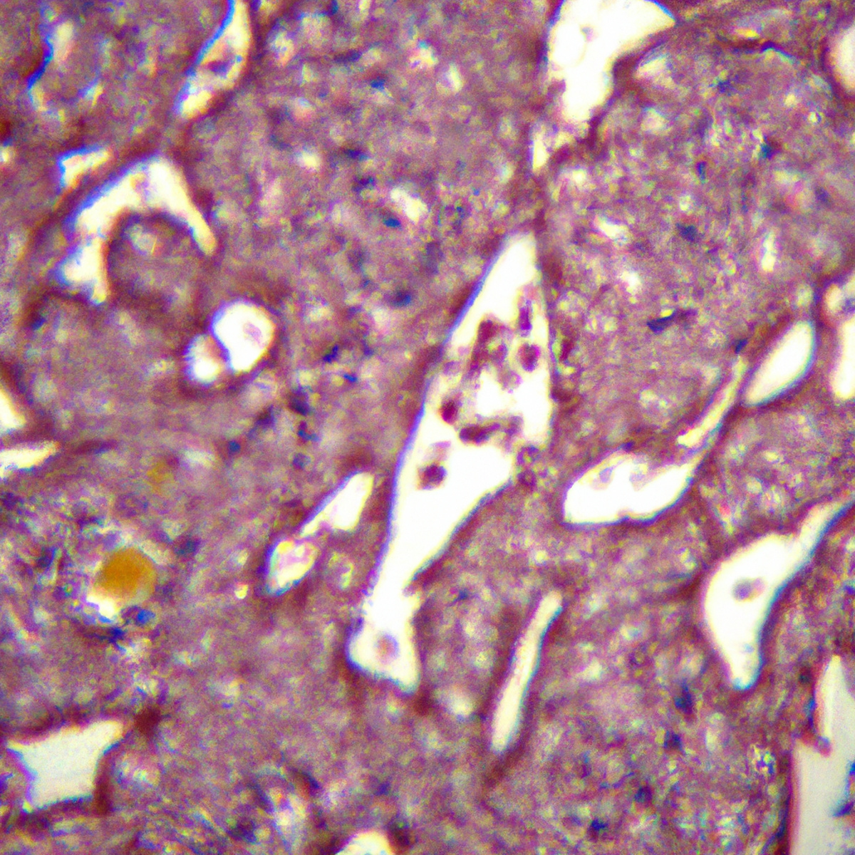
Biopsy and Histopathological Analysis
In some cases, a biopsy of the gallbladder tissue may be necessary to confirm the diagnosis definitively. During a biopsy, a small sample of tissue is collected from the gallbladder and sent for histopathological analysis. This analysis can reveal the characteristic infiltration of inflammatory cells seen in XGC.
Differential Diagnosis
XGC shares symptoms with other gallbladder and gastrointestinal conditions, making a differential diagnosis crucial. Conditions such as acute cholecystitis, gallstones, and malignancies must be ruled out through the diagnostic process.
Conclusion
Xanthogranulomatous Cholecystitis is a rare but serious condition that requires prompt diagnosis and treatment. Recognizing its symptoms, such as abdominal pain, jaundice, fever, nausea, and unexplained weight loss, is essential for early intervention. The diagnosis involves a combination of physical examination, imaging tests, blood tests, and, in some cases, histopathological analysis.
With timely and accurate diagnosis, patients can receive appropriate treatment, which may include surgery to remove the affected gallbladder. If you or someone you know is experiencing symptoms of XGC, seek medical attention promptly to ensure the best possible outcome.